
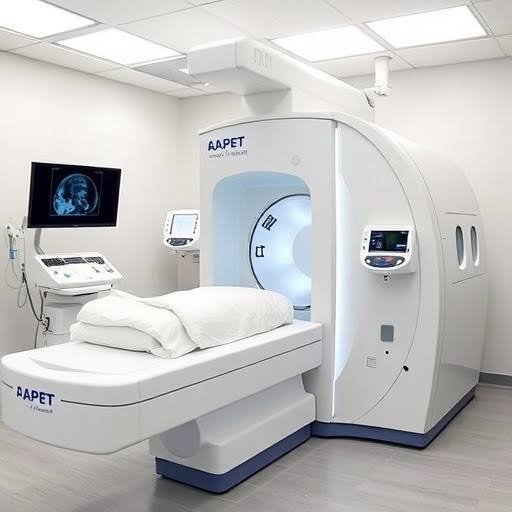
Radiology uses imaging tools to show bones, organs, and soft tissue inside the body, and they use various methods. A radiologist reads the images, and your doctor uses the report to plan next steps. Each technique relies on a different form of energy, which affects what it shows. Here are some of the techniques and technology used in radiology:
Digital X-rays
A digital X-ray sends a small beam of radiation through the body, and a sensor records the result. Since bone absorbs more radiation than soft tissue, it appears white on the image. The image appears on a computer screen within seconds. Radiology teams adjust the contrast and zoom to study small details.
Digital systems typically use less radiation than film because sensors respond to smaller amounts of energy. Doctors order X-rays for fractures, joint changes, chest conditions, and dental exams. The exam takes a few minutes, and you hold still for each image.
Computed Tomography Scans
A CT scanner rotates an X-ray tube around your body. A computer combines the data into cross-sectional slices that show organs, bones, and blood vessels. These slices stack together into 3D views, and radiologists study them from many angles.
Some CT exams use a contrast dye, and the technologist gives it by mouth or through an IV. The dye highlights blood vessels and certain organs on the images. Before the scan, clinics often share steps that patients follow on the day of the exam:
- Avoid food or drink for a few hours if the clinic requests it
- Tell staff about kidney problems or past reactions to contrast dye
- Remove jewelry, eyeglasses, and other metal items
- Wear loose clothing without metal snaps or zippers
Magnetic Resonance Imaging
MRI uses a strong magnet and radio waves instead of radiation, which sets it apart from X-rays and CT. The magnet aligns hydrogen atoms in your body, and radio pulses make them release signals. A computer turns these signals into detailed images.
MRI shows soft tissue with a high level of detail. Doctors order it for brain and spine studies, and they also use it for joint and ligament injuries. Some MRI exams use a contrast agent called gadolinium, which highlights blood flow and inflammation.
The scanner makes loud knocking sounds, so most clinics offer earplugs or headphones. Since the magnet stays on at all times, staff screen every patient for metal before entry. Pacemakers, certain implants, and metal fragments may limit your ability to have an MRI.
Advanced Ultrasound and Fluoroscopy
Ultrasound uses sound waves that bounce off tissues and return to a handheld probe, and it uses no radiation. Advanced systems add Doppler imaging to track blood flow, and 3D ultrasound builds volume images. Fluoroscopy uses a continuous X-ray beam, and the images appear as live video on a monitor. Doctors use it when they need to watch organs or joints in motion, and typical uses include the following procedures:
- Barium swallow studies of the esophagus and stomach
- Joint injections guided by live imaging
- Catheter placement in blood vessels
- Position checks of orthopedic hardware
Visit a Radiology Clinic
Your doctor chooses an imaging test based on your symptoms and medical history. Some exams require a referral, and others need preparation steps such as fasting. When you schedule, ask the radiology clinic which instructions apply to your exam. If you have questions about radiation, contrast dye, or implants, bring them to your appointment.